Basal Cell Carcinoma (abbreviated BCC or BCE)
Basal Cell Carcinoma (abbreviated BCC or BCE for Basal Cell Epithelioma) is the most common type of skin cancer. It is 3–4 times more common than traditional cancers like breast, lung, colon, and others.
When your dermatologist or trained medical professional examines your skin, they are searching for precancerous growths (Actinic Keratoses and Atypical “moles”) and for non-spreading skin cancer like a Basal Cell Carcinoma. They are also checking to be certain that you do not have a more serious skin cancer, such as a Squamous Cell Carcinoma or a Melanoma. Your dermatologist or trained medical professional may use a lighted, magnifying device called a dermatoscope or dermascope to better evaluate your skin. Fortunately, most skin cancers are discovered early, and can be successfully treated.
Remember that you are physically unable to examine many areas of your own body, including your back, buttocks, back of the thighs, and posterior scalp. Your dermatologist or trained medical professional literally “has your back” in searching for skin cancer. A camera phone may enable you to look at some of these areas. Early detection of skin cancer can save your life and that of a family member or friend.
Fortunately, Basal Cell Carcinoma (BCC), the most common cancer of any type, does not normally spread (i.e. metastasize) from its site of origin. However, BCC can enlarge and grow deeply into the skin, which can affect the surrounding skin structures. On the face, a BCC can damage the nose, ears, eyes, and mouth, resulting in partial or complete loss, scars, and disfigurement.
In contrast, the second most frequent skin cancer, Squamous Cell Carcinoma, can spread into your bloodstream (metastasize). The third most frequent skin cancer, Melanoma, is considered the most dangerous of all types of cancer due to its easy ability to metastasize.
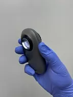
Dermatoscope
A special, lighted magnifying instrument is used to see your “moles” more clearly
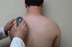
Dermatoscope Exam
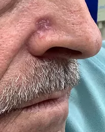
Basal Cell Carcinoma
Right nasal fold
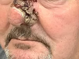
Basal Cell Carcinoma
Skin cancer can lead to local destruction of the nose, ear, lips, and other body areas.
This photo was taken BEFORE any surgery.
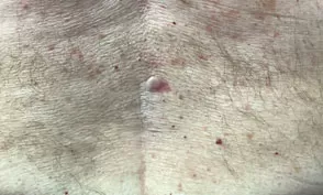
Basal Cell Carcinoma, mid-back
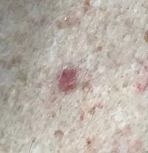
Basal Cell Carcinoma, mid-back
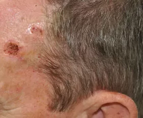
Basal Cell Carcinoma
Two BCCs are seen on the left upper forehead.
These usually only invade locally and rarely spread elsewhere in the body (i.e. metastasize).
RISK FACTORS FOR BASAL CELL CARCINOMA
Skin cancer occurs in all races, regardless of their skin color. It is more common in fair-skinned individuals. See if you or your family have any of these Risk Factors:
[Resources from the American Academy of Dermatology and the American Society for Dermatologic Surgery were consulted for this section.]
DIAGNOSIS OF BASAL CELL CARCINOMA
Your dermatologist or trained medical professional will examine your skin to look for suspicious growths. Basal cell carcinomas can have a variety of appearances, from a white or pink, translucent “bump” to a draining, non-healing “sore”. Basal Cell Carcinoma may also appear as a pink scar or even a brown spot.
To diagnose Basal Cell Carcinoma (BCC), your dermatologist or trained medical professional can perform a skin biopsy under local anesthesia at the time of your office visit. This is the only way to confirm the diagnosis of skin cancer. A skin biopsy procedure is fast, safe, and usually heals with minimal scarring.
If you have a suspicious growth, a small sample of skin is surgically removed (i.e. biopsied) under local anesthesia. This specimen is sent to a pathology laboratory for processing and examination under a microscope by a pathologist. A pathology report is usually available within 7-14 days. Your dermatologist or medical care professional will discuss your pathology report with you at the time of your next doctor’s visit. If a skin cancer diagnosis is found on your pathology report, treatment options will be discussed with you.
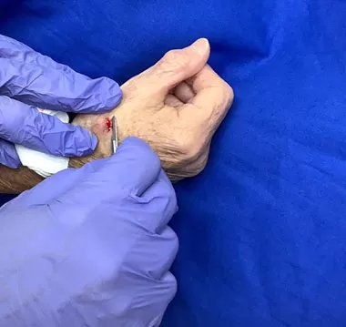
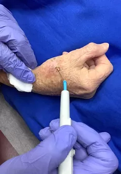
A growth that does not heal or keeps recurring should be biopsied. After local anesthesia, a shave or scallop biopsy is performed with a scalpel and blade. The biopsy site is then “burned” (electrodessicated) with the probe of an electrosurgery unit. The specimen is sent out for pathology.
A skin biopsy is usually covered by Medicare, Medicaid, and insurance companies, minus any deductible or co-payment required by the insurer. If you have a second insurance policy (“secondary insurance”), this will usually cover your co-payment.
TREATMENT OPTIONS FOR BASAL CELL CARCINOMA
Even though Basal Cell Carcinomas (BCC) rarely ever spread elsewhere (i.e metastasize), they can grow deeply into your skin, causing local tissue destruction. This may lead to the partial or complete loss of your nose, ears, lips, or eyes if not treated early on. Basal Cell Carcinomas can also cause scarring and other types of disfigurement. Your dermatologist or medical care professional can help you decide on the best treatment for your Basal Cell Carcinoma.
Treatment options for Basal Cell Carcinoma (BCC) depend on the location, size, type of BCC, and age of the patient. Your dermatologist or trained health care profession must consider many different conflicting factors. For example, many patients are sensitive to pain, do not like needles, or are taking blood thinners. These patients may prefer Superficial Radiation Therapy (SRT) [see (8)], as there are no needles, no pain, no risk of bleeding from blood thinners, and no interference with one’s lifestyle.
Other treatment factors are the location and type of skin cancer. If the base of the BCC is less than 1.0 cm (0.4 inches) in size, it is considered low risk; over 1.0 cm in size, it is high risk. If the BCC is located near an eye, ear, nose, mouth, or temple area, it is considered high-risk. A Superficial BCC is easily treated, while a sclerotic / fibrotic BCC may be larger than it appears and requires excision or Mohs surgery [see (5)]. A less invasive procedure such as Superficial Radiation Therapy (SRT) [see (8)] may be preferred for elderly patients and for those with severe disabilities.
SURGICAL TREATMENTS
(1) Electrodessication and Curettage for Basal Cell Carcinoma
This is one of the most common ways to treat non-melanoma skin cancers like Basal Cell Carcinoma and Squamous Cell Carcinoma. This is sometimes abbreviated ED&C or EDC or EC or CE (electrodessication and curettage), F&C or FC (fulguration and curettage). Electrodessication is when the electrosurgery probe touches the skin area during treatment while fulguration is when the probe is allowed to spark over the treatment area.
This treatment consists of local anesthesia followed by “burning” the skin with an electrosurgery probe (i.e., electrodessication and/or fulguration), then scraping the skin cancer area with a curette. This electrosurgery-curettage technique is usually repeated two or three times to destroy the underlying skin cancer. It works best for skin cancers that are small (i.e., less than 1.0 cm (0.4 inches) in
size.
This treatment is usually covered by Medicare, Medicaid, and insurance carriers minus any deductible or co-payment required by your insurer. If you have a second insurance policy (“secondary insurance”), this will usually cover your co-payment.
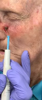
Electrodessication
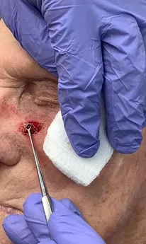
Curettage
Skin cancer is often treated with electrodessication (“burning” the skin cancer with the probe of an electrosurgery device), followed by curettage (scraping the base of the skin cancer with a curette). This is usually repeated 2–3 times.
(2) Excision of Basal Cell Carcinoma
Your dermatologist or medical professional can “cut out” (i.e., excise) your skin cancer under local anesthesia using a scalpel. The excised skin cancer is sent to a pathologist for processing and examination under a microscope to be certain that the skin specimen margins are free of skin cancer. Excision is usually performed on skin cancer that is over 1.0 cm (0.4 inches) in size. An area around the skin cancer of about 0.4 cm (0.2 inches) to 1 cm (0.4 inches) is usually required to completely excise (cut out) the skin cancer. Some skin cancer excisions are larger and may require moving normal skin over the cancer treatment site (skin flap) or detaching nearby skin to cover the surgery site (skin graft).
Excisional treatment is covered by Medicare, Medicaid, and insurance
carriers, minus any deductible or co-payment required by your insurer. If you have a second insurance policy (“secondary insurance”), this will usually cover your co-payment.


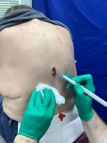
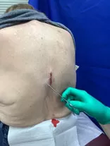
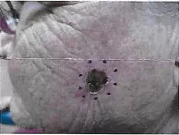
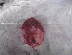
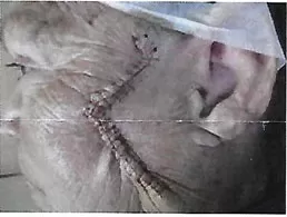
An atypical growth may be excised under local anesthesia. A drawing is made around the skin cancer, as seen in Photo 1. The size of the drawing is normally 4–10 mm around the growth to be certain that all of it is removed. In Photo 2, the growth is being excised (cut out), followed by stopping any bleeding with electrosurgery (heat applied by the probe of an electrosurgery device), as seen in Photo 3. The final step is to tie the skin together with sutures (stitches), as seen in Photo 4.
(3) Laser Treatment for Basal Cell Carcinoma
A laser is sometimes used to treat superficial skin cancer. Your dermatologist or medical professional may use a focused beam of light (laser) on an area under local anesthesia to destroy superficial non-melanoma skin cancers, Basal Cell Carcinoma and Squamous Cell Carcinoma.
Medicare, Medicaid, and insurance carriers may cover laser treatment for skin cancer minus any deductible or co-payment required by your insurer. If you have a second insurance policy (“secondary insurance”), this will usually cover your co-payment.
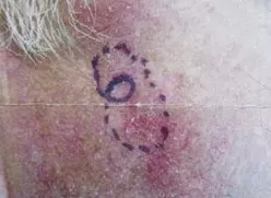
BEFORE Laser Treatment for non-melanoma skin cancers (e.g. Basal Cell and Squamous Cell Carcinoma)
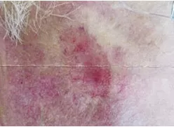
AFTER Laser Treatment: Crusting of the treatment site
(4) Cryosurgery for Basal Cell Carcinoma
Cryosurgery treatment is occasionally used to treat skin cancer. Cryosurgery itself can cause scarring and, occasionally, disfigurement. Some dermatologists use cryosurgery for Superficial Basal Cell Carcinomas (BCC) and Squamous Cell Carcinomas (SCC) in elderly patients and in resistant BCCs and SCCs which have ill-defined borders or depth. Mohs surgery [see (5)] or Superficial Radiation Therapy (SRT) [see (8)] are usually preferred over cryosurgery.
This type of cryosurgery treatment consists of spraying liquid nitrogen in two freeze-thaw cycles of one minute each. The treated areas will become frozen and turn white. After two freeze-thaw cycles, the white color fades over several minutes. The treatment site then becomes red and swollen, and it may form a draining wound over several days or weeks. The area will take 4–6 weeks or longer to heal. For the best cosmetic result, gently cleanse the treatment site twice a day with mild soap and water, followed by the application of Vaseline or Aquaphor.
Cryosurgery for skin cancer is normally covered by Medicare, Medicaid, and insurance companies, minus any deductible or co-payment required by your insurer. If you have a second insurance policy (“secondary insurance”), this will usually cover your co-payment.
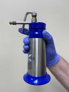
Cryosurgery Unit
Cryosurgery is commonly used to treat precancerous growths (Actinic Keratoses).
Cryosurgery is occasionally used to treat non-melanoma skin cancer (Basal Cell Carcinoma and Squamous Cell Carcinoma).
(5) Mohs surgery for Basal Cell Carcinoma
Mohs surgery is named after Fredric E. Mohs (1910-2002), who pioneered this procedure. This is considered the “Gold Standard” for skin cancer treatment. Mohs surgery is generally performed on high-risk areas around the eyes, ears, nose, or mouth and for recurrent skin cancer. There is up to a 99% cure rate with Mohs surgery, which is superior to most other forms of skin cancer treatment. Superficial Radiation Therapy (SRT) [see (8)] also has a very high cure rate of 98–99%.
If you are referred to a Mohs surgeon, the Mohs surgeon will evaluate your skin cancer site. Your skin cancer will be removed in stages under local anesthesia, usually the same day. A layer of skin is removed in each stage, and the tissue examined under a microscope at an on-site laboratory. This is repeated until the skin cancer has been completely removed.
During your office visit to a Mohs surgeon, you will have to wait between each stage of your skin treatment for your skin specimen to be processed and evaluated under a microscope. Plan for your visit to a Mohs surgeon to take several hours or longer.
You may also be required to stay longer for reconstructive surgery after Mohs surgery if there is a large surgical wound created by your Mohs surgery treatment. Reconstructive surgery may include complex repair, a skin graft, or a skin flap. Alternatively, the wound from Mohs surgery may be repaired at a later date or may be allowed to heal naturally over 4-6 weeks.
Medicare, Medicaid, and insurance carriers generally pay for Mohs surgery and Superficial Radiation Therapy (SRT) minus any deductible or co-insurance required by your insurer. If you have a second insurance policy (“secondary insurance”), this will usually cover your co-payment.
Before Mohs Treatment for a Non-Melanoma Skin Cancer (Basal Cell or Squamous Cell Carcinoma).
Excision (cutting out) of Non-Melanoma skin cancer (i.e. Basal Cell or Squamous Cell Carcinoma). Note That the size of the skin cancer repair is much larger than expected.
After surgical repair of the Mohs defect with an advancement flap.
NON-SURGICAL TREATMENT FOR BASAL CELL CARCINOMA
(6) TOPICAL TREATMENT FOR BASAL CELL CARCINOMA
Your dermatologist or medical professional may also use a topical
anti-cancer cream, gel, or solution for treatment of small Superficial Basal Cell Carcinomas. Superficial Basal Cell carcinomas are on the skin surface and have not invaded deeper into the skin. Topical anti-cancer creams are able to reach these superficial lesions. Other types of Basal Cell Carcinoma, Squamous Cell
Carcinoma, and Melanoma are deeper, where the anti-cancer cream may not reach. Consequently, the surface of the skin cancer may heal, while the deeper part of the skin cancer can enlarge and spread (metastasize).
Alternatively, an anti-cancer cream, gel, or solution may be used after previous skin cancer treatment in an effort to prevent skin cancer recurrence.
5-Fluorouracil (Eudex™, Tolak™) and Imiquimod Cream (Aldara™) are commonly used to treat small, Superficial Basal Cell Carcinomas. They are usually applied 5–7 days a week for 6–8 weeks or longer. Treated areas will become red, swollen, and may crust over before healing about 3–4 weeks after discontinuing the anti-cancer cream therapy. Patients must avoid getting the anti-cancer medications in their eyes, as they can cause eye irritation or damage.
A new alternative anti-cancer medication is Klisyri™ (tiranibulin) ointment. Currently, Klisyri™ is only approved for the treatment of precancerous Actinic Keratoses. This ointment is applied daily for five days to pre-cancerous areas while avoiding eye areas. The treated areas become red, swollen and may crust over before healing in 2–4 weeks.
It is currently unknown whether Klisyri™ will help Superficial Basal Cell Carcinoma or other types of skin cancer. Any treatment in the future will probably involve several treatment courses. Klisyri™ ointment is expensive unless covered by a discount coupon or by filing your prescription directly with a specialty pharmacy through your doctor’s office.
Medicare, Medicaid, and insurance companies usually cover the office visit charges for the treatment of skin cancer minus any deductible or co-payment required by your insurer. If you have a second insurance policy (“secondary insurance”), this will usually cover your co-payment. Insurance companies may not want to cover or delay coverage of your anti-cancer cream. This problem can be avoided by filing your prescription directly with a specialty pharmacy through your doctor’s office.
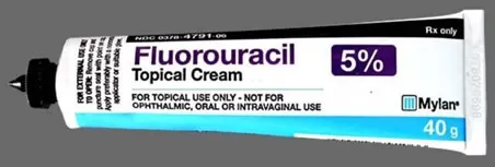
5-Fluorouracil Cream (Efudex™, Tolak™) is commonly used to treat precancerous areas (Actinic Keratosis) and Superficial Basal Cell Carcinoma. It is usually prescribed for 5-7 days a week for 6–8 weeks or longer for treatment of Superficial Basal Cell Carcinoma.
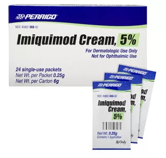
Imiquimod Cream Aldara™ is sometimes used to treat precancerous skin lesions (Actinic Keratoses) and Superficial Basal Carcinoma. The cream usually comes in packets, and a small amount is applied to the affected areas. Imiquimod Cream is usually prescribed 5-7 days a week for 6–8 weeks or longer for treatment of Superficial Basal Carcinomas.
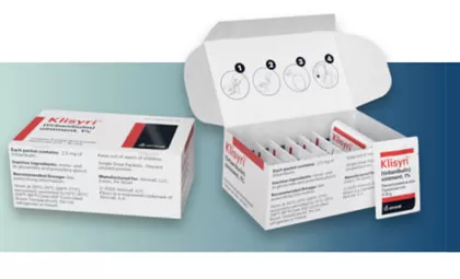
Klisyri ointment is a new treatment for precancerous skin growths (Actinic Keratoses). Klisyri is usually applied once a day for five days for treatment of Actinic Keratoses. It is unknown at this time whether Klisyri will help with superficial basal cell carcinomas or other types of skin cancer.
(7) BLUE OR RED LIGHT TREATMENT FOR BASAL CELL CARCINOMA (also known as PHOTODYNAMIC THERAPY PDT)
Blue or Red Light Therapy is sometimes used to treat single or multiple, superficial non-melanoma skin cancers (e.g. Basal Cell Carcinoma and Squamous Cell Carcinoma.) This therapy should not be used for large or deep skin cancers or for Melanoma.
Therapy consists of the application of a special solution or gel to the treatment area at your doctor’s office. The solution or gel gets into the “nooks and crannies” of your skin and makes the skin sensitive to Blue and Red light. After waiting 1 hour for the face or scalp, or 2–3 hours for other body areas, the Blue Light Treatment is administered by a light stand for 10 to 16.5 minutes. Alternatively, Red Light Therapy can be administered with a smaller light stand. Red light from the smaller light stand is presently given for 10 minutes to 2 adjacent treatment areas (a total of 20 minutes). A larger Red Light Therapy light stand is pending approval.
After the Blue or Red Light Treatment, your skin becomes red and slightly swollen. You should avoid sunlight and protect yourself from the sun for the next 24-48 hours, as you may have become sun-sensitive. Your skin will usually heal within a few weeks and be much softer and smoother. Repeat therapy every month for several months may be required, depending on how your Basal Cell Carcinoma responds to Blue or Red Light therapy.
Medicare, Medicaid and Insurance plans may cover Blue or Red light therapy for Basal Cell Carcinoma minus any deductible or co-payment required by the insurer. If you have a second insurance policy (“secondary insurance”), this will usually cover your co-payment.
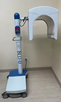
Blue Light Unit
Blue and Red Light treatments are used to treat sun-damaged skin and precancerous lesions (Actinic Keratoses)
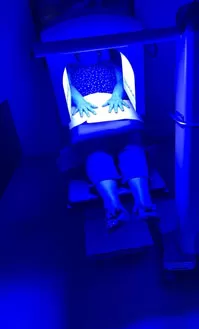
Blue Light Treatment
Patient receiving Blue Light Treatment to the Arms for Actinic Keratoses
(8) SUPERFICIAL RADIATION THERAPY (SRT) FOR BASAL CELL CARCINOMA
Radiation therapy has been used to treat skin cancer since 1896. Treatment is based on Wilhelm Rontgen's remarkable discovery of X-rays on November 8, 1895. Over the last century, radiation therapy has evolved. In the past 8 years, safe, reliable radiation machines have been developed that use tungsten instead of radioactive materials to treat Basal Cell and Squamous Cell Carcinomas.
Superficial Radiation Therapy (SRT) may be the preferred treatment method for patients who are afraid of needles or take blood thinners. SRT may also be the best choice for elderly patients or for patients with severe disabilities.
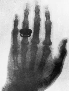
The first X-ray of a human hand on December 22, 1895. This X-ray is of Wilhelm Rontgen’s wife’s hand.
Sensus Healthcare™ has been a pioneer in radiation therapy and manufactures the Sensus SRT 100™, 100+™, and Vision™ radiation machines for skin cancer treatment. SRT uses superficial radiation which does not go deeper than your skin. In contrast, hospital radiation machines penetrate deep below the skin surface to treat breast cancer, colon cancer, prostate cancer, and other internal cancers. For this reason, superficial radiation therapy (SRT) has minimal side effects in contrast to the side effects of hospital radiation machines.
Several other companies have recently developed radiation therapy for non-melanoma skin cancer. Comparison studies are not presently available.
The Advantages of Superficial Radiation Therapy (SRT) for Basal Cell Carcinoma and Squamous Cell Carcinoma are:
SRT therapy is usually not used on children or patients with genetic disorders like basal cell nevus syndrome, xeroderma pigmentosum, albinism, or rare skin cancers that are resistant to radiation therapy. Mohs surgery [see (5)] is the preferred treatment for these less common cases.
Superficial Radiation Therapy (SRT) is covered by Medicare, Medicaid, and insurance carriers, minus any deductible or co-payment required by your insurer. If you have a second insurance policy (“secondary insurance”), this will usually cover your co-payment.
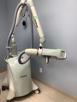
Sensus SRT 100+ Radiation machine Superficial radiation machines are very safe. They produce radiation by the use of tungsten. This is the same element that is found in your electric light bulb.
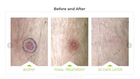
Treatment for Non-Melanoma Skin Cancer (Basal Cell and Squamous Cell Carcinomas) with Superficial Radiation Therapy (SRT).
TREATMENT FOR ADVANCED BASAL CELL CARCINOMA
Basal Cell Carcinoma rarely becomes invasive or spreads (metastasizes). There are several new drug therapies that may shrink or help control aggressive Basal Cell Carcinoma:
These anti-cancer drugs can be used before surgery to shrink an advanced Basal Cell Carcinoma or after surgery on a Basal Cell Carcinoma resistant to surgery. Odomzo™ or Erivedge™ are commonly prescribed by your dermatologist. Your skin cancer treatment is monitored during regular dermatology visits.
These cancer treatments are covered by Medicare, Medicaid, and other insurance carriers minus your deductible or co-payment required by your insurance. A second insurance policy (“secondary insurance”) will usually cover your co-payment.
